1
/
of
1
TecAfrica Solutions
DW-P60(P8Lite) Best Portable medical cardiac ultrasound scanner machine(Echo machine)
DW-P60(P8Lite) Best Portable medical cardiac ultrasound scanner machine(Echo machine)
Taxes included.
Shipping calculated at checkout.
Pickup available at FANCOURT OFFICE PARK
Usually ready in 2-4 days
DW-P60 Portable Cardiac medical Color Doppler Ultrasound System
Based on Dawei's new generation ultrasound platform-4.0S, P60 medical ultrasound machine has raised the industry standards to an all new level. Advanced signal transmission and reception processors provide highly sensitive and accurate echo detection. Innovative transducer technologies allow for better penetration, higher resolution, greatly enhancing your diagnostic experience.
-
Abdomen
Vascular
Cardiology
OB & GYN
Urology
Musculoskeletal
Interventional ultrasound
Small parts
Anesthesiology
Pediatrics
![P60 [已恢复]-01](https://www.ultrasounddawei.com/uploads/P60-%E5%B7%B2%E6%81%A2%E5%A4%8D-01.png)
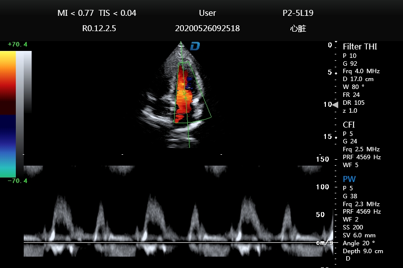
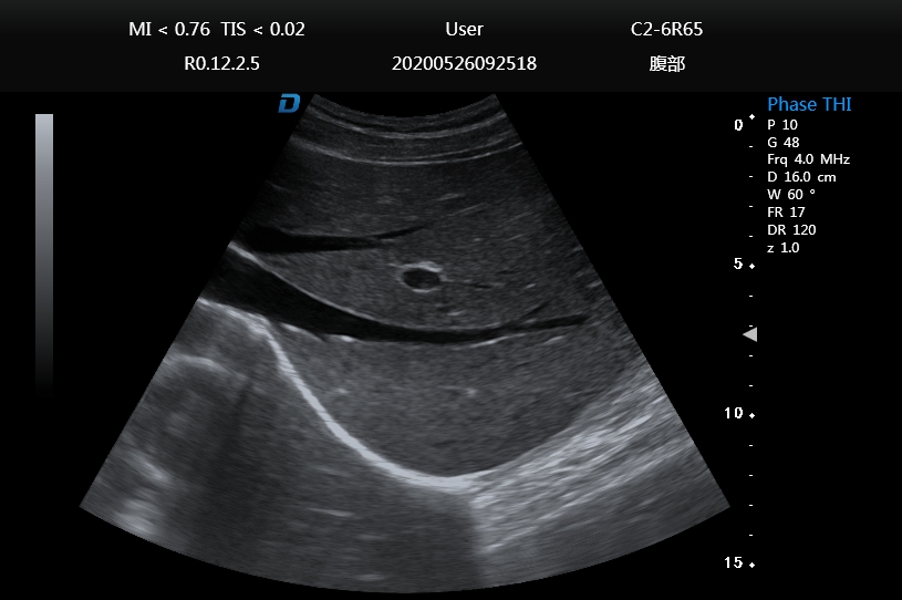
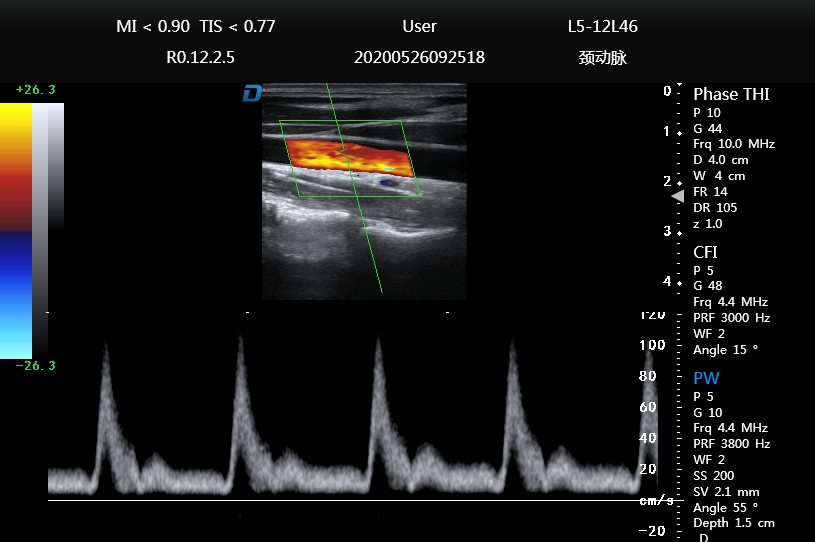
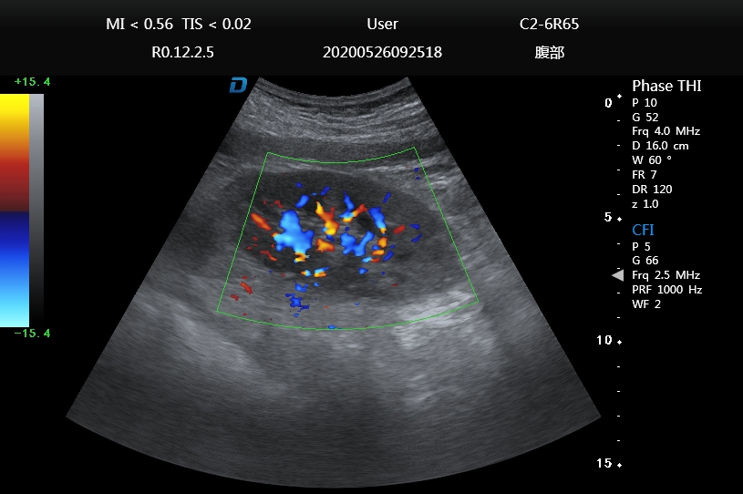
High Definition
And High Quality
More powerful to have powerful image processing functions, a
wide range of technical applications, harmonic imaging, harmonic fusion,
free arm 3D, elastography, trapezoidal imaging, contrast imaging.
Trapezoidal imaging
Refers to converting the line data of a linear
array probe into a trapezoidal image through
coordinate transformation and interpolation,
which is an extended imaging.
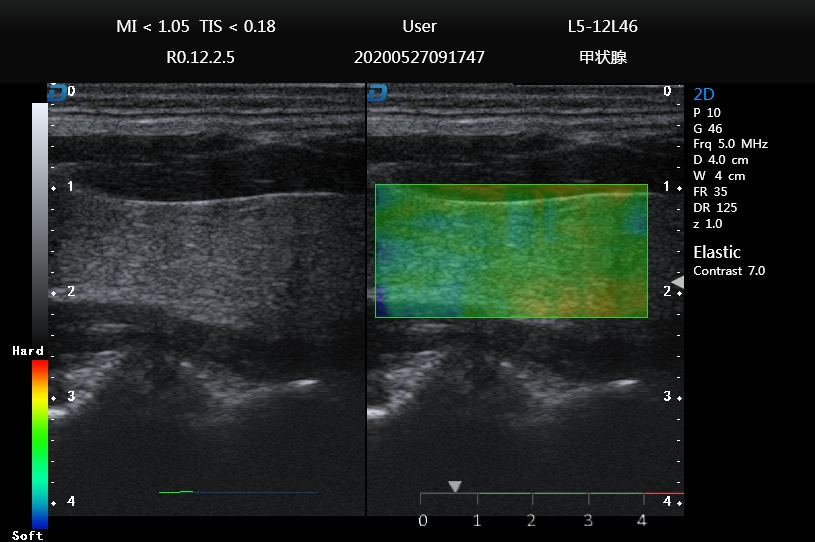
Freehand elastography mode
Freehand elastography can help doctors
distinguish between soft/hard lesions
and surrounding tissues.
Wide-field imaging
Also known as ultra-wide-field imaging.
Compared with ordinary ultrasound imaging,
wide-field imaging provides a new perspective
for clinical diagnosis, and has a very important
clinical significance for observing large lesions
and the relationship between the lesions and
surrounding tissues. Diagnostic significance.
Contrast imaging
The use of significantly different echo charac
teristics from human soft tissue, or acoustic
characteristic impedance (i.e., specific acous
tic impedance).
Freehand 3D imaging mode
Provides a method for generating 3D images when using standard linear array, convex array,
and cavity probe inspection. The process of freehand 3D imaging is to obtain a series of frame
images (refer to the below figure, move the probe in parallel at a uniform speed) apply volume
rendering technology to reconstruct the volume data and display the 3D rendered image.
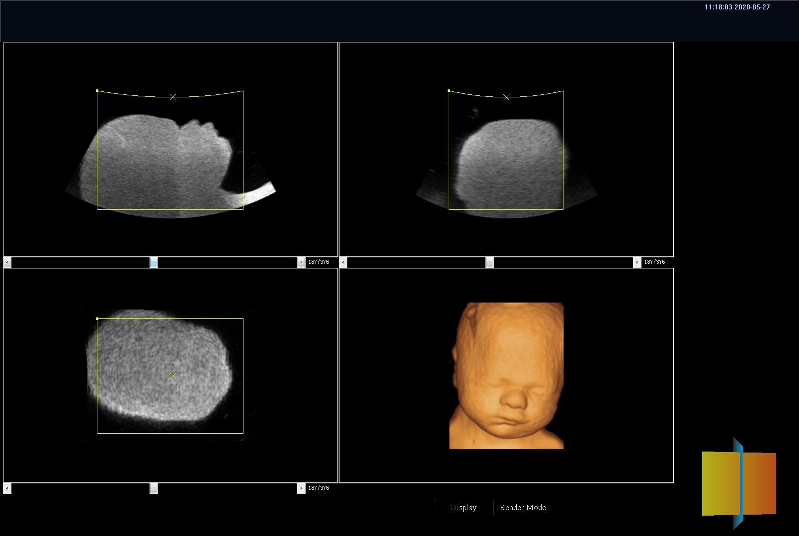
Volume 3D / 4D imaging mode
4D Imaging, also known as real-time 3D imaging, provides an interactive
means of viewing dynamic 3D imaging. The freehand 3D imaging mode has
different probe movement speeds, during 4D imaging inspection, the volume
probe is fixed at one location and cannot be moved. The machinery inside
the probe the component can perform stable continuous scanning of different
positions by swinging, so as to obtain a series of continuous and stable
frame images. It can be seen that the quality of the 4D rendered image is
significantly higher than the 3D imaging with the bare hand.
Anatomical M-mode
Has only one m-sampling line, which has
limitations for moving examination tissues,
especially for difficult patients. The anatomical
M-mode makes up for the lack of traditional
M-mode for the examination of patients with
difficult imaging, and it provides multiple
M-sampling lines. To enable you to perform
more effective motion analysis on M-mode
images at different angles and positions.
Puncture enhancement
Automatic detection of the needle body,
automatic deflection of the sound beam, and
smart puncture enhancement technology make
the puncture display in the human body more
intuitive.
Tissue doppler imaging mode
Abbreviated as TDI mode, using Doppler principle to estimate tissue motion, such as the speed of myocardial
motion.The TDI mode can obtain motion information and generate color-coded images of tissue motion speed.
Automatic IMT measurement
The thickness of the intima of the blood vessel is an important indicator for predicting the
risk of cardiovascular disease. The automatic measurement technique of the intima can
automatically measure the thickness of the intima in the near and far fields of the blood
vessel and automatically optimize the measurement angle.


